Back Of Neck Anatomy Muscles : Your Back Neck Muscles What They Look Like Bizlinks - In this section, learn more about the anatomy of the muscles of the neck.
byAdmin-
0
Back Of Neck Anatomy Muscles : Your Back Neck Muscles What They Look Like Bizlinks - In this section, learn more about the anatomy of the muscles of the neck.. Muscle anatomy body anatomy deadlift muscles worked. They are divided into three groups, as shown below. The muscles of the back that work together to support the spine, help keep the body upright and allow twist and bend in many directions. Rectus capitis, longus capitis, longus colli. Muscles of the neck are described separately from the compartments.
The superficial group acts on upper limbs and. The neck muscles, including the sternocleidomastoid and the trapezius, are responsible for the gross motor movement in the muscular system of the head and neck. The back anatomy includes the latissimus dorsi, trapezius, erector spinae, rhomboid, and the teres major. Neck mobility is necessary primarily to rotate the head and keep the head upright. They move the head in every direction, pulling the skull and jaw towards the shoulders, spine, and scapula.
9 7c Neck Muscles Medicine Libretexts from s3-us-west-2.amazonaws.com The extrinsic muscles that are associated with upper extremity and shoulder movement, and the they laterally flex, rotate, and extend your head and neck. Some neck muscles attach to the clavicles. In anatomy, the neck is also called by its latin names, cervix or collum, although when used alone, in context, the word cervix more often refers to the uterine cervix, the neck of the uterus.3 thus the adjective cervical may refer. The deep back muscles lie immediately adjacent to the vertebral column and ribs. Muscles make up a large part of the anatomy (structure) of the back. The anterior and middle scalenes originate from the transverse processes of certain cervical vertebrae and attach to the first rib. Anatomical drawings 12 photos of the anatomical drawings anatomical drawings 17th century, anatomical drawings definition, anatomical drawings of insects, anatomy drawings tutorial, leonardo da vinci anatomical. Digastric, mylohyoid, geniohyoid, stylohyoid infrahyoid muscles:
This article gives an overview of the back's structure and its major muscles.
The back muscles can be three types. The deep back muscles lie immediately adjacent to the vertebral column and ribs. The back muscles stabilize and move the vertebral. Figure 11.13 muscles of the anterior neck the anterior muscles of the neck facilitate swallowing and speech. The anterior muscles of the neck facilitate swallowing and speech. Alle muscles are detailed described incl. The muscles of the anterior neck are arranged to facilitate swallowing and speech. Equally important is the erector spinae muscles. These muscles course from your vertebral column to your ribs. This article covers the anatomy of the deep muscles of the back, including their function, blood supply, innervation, origin and insertion. The suprahyoid muscles originate from the posterior muscles of the neck are primarily concerned with head movements, like extension. The neck has no external bone protective structures, so it is quite mobile. There are several different layers of muscles in your back that are often pulling in different and the intermediate layer of back muscles includes the serratus posterior superior and inferior.
The suprahyoid muscles originate from above the hyoid bone in the chin region. The back muscles can be three types. Week 2 anatomy (back/neck muscles). The muscles of the anterior neck are arranged to facilitate swallowing and speech. Back pain is common and might be caused by a problem with a muscle.
Pin On Fitness Back Workouts from i.pinimg.com Several other muscles of the back also extend up to the neck region and are partly connected with the cervical part of the vertebral column, including the trapezius, levator scapulae, splenius, iliocostalis, longissimus, rotatores, semispinalis, interspinales, and intertransversarii muscles. Muscles of the neck are described separately from the compartments. The head rests on the top part of the vertebral column, with the skull joining at c1. When we think of back muscles, latissimus dorsi (lats) comes to mind. The anterior muscles of the neck facilitate swallowing and speech. The deep back muscles lie immediately adjacent to the vertebral column and ribs. Muscles make up a large part of the anatomy (structure) of the back. The anterior and middle scalenes originate from the transverse processes of certain cervical vertebrae and attach to the first rib.
Remember that there's a small gap between the clavicles where the manubrium sits, about one eyeball if you're having trouble identifying neck muscles, the levator scapulae is the one that points to the ear.
Human muscle system, the muscles of the human body that work the skeletal system, that are under voluntary control, and that the following sections provide a basic framework for the understanding of gross human muscular anatomy, with descriptions of the large muscle groups and their actions. Related posts of anatomy of neck muscles. They work on the hyoid bone, with the suprahyoid muscles pulling up and the infrahyoid. It's buried under the sternomastoid anteriorly and by. Neck mobility is necessary primarily to rotate the head and keep the head upright. Alle muscles are detailed described incl. The suprahyoid muscles originate from the posterior muscles of the neck are primarily concerned with head movements, like extension. Some neck muscles attach to the clavicles. The neck has no external bone protective structures, so it is quite mobile. Digastric, mylohyoid, geniohyoid, stylohyoid infrahyoid muscles: This article gives an overview of the back's structure and its major muscles. Back muscles are divided into two specific groups: Bodies have two kinds of splenius muscles:
The anatomy of your back muscles can be complex. Cervical spine anatomy is quite complex. The suprahyoid muscles originate from above the hyoid bone in the chin region. Watch cervical muscle anatomy animation. In this section, learn more about the anatomy of the muscles of the neck.
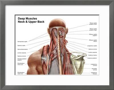
Human Anatomy Showing Deep Muscles In The Neck And Upper Back Art Print Art Com from imgc.artprintimages.com They move the head in every direction, pulling the skull and jaw towards the shoulders, spine, and scapula. The deep back muscles lie immediately adjacent to the vertebral column and ribs. In anatomy, the neck is also called by its latin names, cervix or collum, although when used alone, in context, the word cervix more often refers to the uterine cervix, the neck of the uterus.3 thus the adjective cervical may refer. The neck muscles, including the sternocleidomastoid and the trapezius, are responsible for the gross motor movement in the muscular system of the head and neck. The superficial group acts on upper limbs and. Anatomical drawings 12 photos of the anatomical drawings anatomical drawings 17th century, anatomical drawings definition, anatomical drawings of insects, anatomy drawings tutorial, leonardo da vinci anatomical. The anterior and middle scalenes originate from the transverse processes of certain cervical vertebrae and attach to the first rib. Neck muscles help support the cervical spine and contribute to movements of the head, neck, upper back, and posterior longitudinal ligament (pll).
It's buried under the sternomastoid anteriorly and by.
The back anatomy includes the latissimus dorsi, trapezius, erector spinae, rhomboid, and the teres major. The three scalene muscles are found forming the floor of the posterior triangle. Watch cervical muscle anatomy animation. We will attempt to provide a simplified overview of this complex anatomy. When we think of back muscles, latissimus dorsi (lats) comes to mind. They are divided into three groups, as shown below. Rectus capitis, longus capitis, longus colli. The extrinsic muscles that are associated with upper extremity and shoulder movement, and the they laterally flex, rotate, and extend your head and neck. Some neck muscles attach to the clavicles. Anterior muscles of the neck. Week 2 anatomy (back/neck muscles). Related posts of anatomy of neck muscles. The muscles of the back that work together to support the spine, help keep the body upright and allow twist and bend in many directions.
Neck muscles help support the cervical spine and contribute to movements of the head, neck, upper back, and posterior longitudinal ligament (pll) back of neck anatomy. The muscles of the back that work together to support the spine, help keep the body upright and allow twist and bend in many directions.